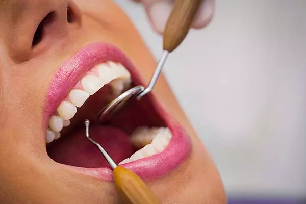
Procedures
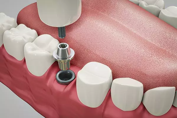
DENTAL IMPLANTS
The teeth are formed by the crown, which is the visible part of the tooth and the root which goes in the jaw bone both form a unit that allows one to chew. Dental Implants are titanium posts designed to replace the missing root of the tooth. After the placement of the implant a healing period is necessary, then a post and a crown(tooth) is placed to fully recover the mastication or chewing abilities, teeth estethics and self confidence. Implants will also prevent the natural bone loss due to missing teeth and can also be placed in case of broken teeth that cannot be saved.
Know about Dental Implants Surgery >>
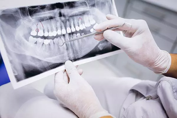
EXTRACTION AND IMMEDIATE DENTAL IMPLANTS PLACEMENT
Extraction of the tooth and implant placement during the same procedure.
Before the immediate implant procedure was developed patients waited at least 8 to 12 weeks for the extraction area to heal in order to receive a dental implant, but because of the constantly changing dental implant and restorative fields it is no longer necessary to wait for the extraction site to heal.
In some cases and after evaluation by the Doctor an immediate implant may be indicated which is a less invasive treatment and will also save you time to have your crown (tooth) sooner. If your bone is adequate both in quantity and quality you may be a candidate to have a temporary tooth placed the same day.
Know about Surgical Instructions | Extraction and Dental Implants placement>>
EXTRACTION AND IMMEDIATE
DENTAL IMPLANTS PLACEMENT
Before the immediate implant procedure was developed patients waited at least 8 to 12 weeks for the extraction area to heal in order to receive a dental implant, but because of the constantly changing dental implant and restorative fields it is no longer necessary to wait for the extraction site to heal.
In some cases and after evaluation by the Doctor an immediate implant may be indicated which is a less invasive treatment and will also save you time to have your crown (tooth) sooner. If your bone is adequate both in quantity and quality you may be a candidate to have a temporary tooth placed the same day.
Know about Surgical Instructions | Extraction and Dental Implants placement>>
IMPACTED WISDOM TEETH
(THIRD MOLARS)
Wisdom teeth are the last teeth to erupt in the mouth. They usually start to come out after 16 or 17 years old, if the teeth erupt into the mouth properly and gum tissue is healthy, wisdom teeth do not have to be removed. This may not be always the case and they may grow inclined, angulated or may be impacted under the gum or bone. When the wisdom teeth are not properly aligned or are impacted or partially erupted they can cause many problems like pain, swelling, infection, difficulty swallowing, pressure and shifting of the teeth. Cyst and tumors may develop around the wisdom teeth and the bone may be destructed or the teeth may be damaged. Extraction of the wisdom tooth and excision of the cyst usually resolves these problems. The surgery to remove the wisdom teeth is done in the oral surgeon office under local anesthesia, laughing gas or IV sedation.
FACIAL TRAUMA
The Oral and Maxillofacial Surgeon is trained and qualified to manage the emergency care, acute treatment, long term reconstruction and rehabilitation of facial trauma and most injuries to the face including jaw fractures, frontal bone, nose, eye socket and cheek bone fractures, lacerations (cuts) to lips and face structures including nerves and knocked out teeth. The Oral and Maxillofacial Surgeons are affiliated and deliver emergency room coverage in local hospitals for facial injuries. These injuries caused by falls, motor vehicle accidents, accidental falls, sports injuries, interpersonal violence, and work-related injuries, may affect patient’s facial appearance, in order to repair such injuries the facial bones are exposed through small well hidden incisions and in the case of jaw fractures the incisions are made inside the mouth avoiding scars on the face. Injuries to teeth are common. Oral surgeons are involved in treating fractures in the supporting bone or in reimplanting and splinting teeth that have been displaced or knocked out.
PRE- PROSTHETIC SURGERY
The surgery performed on your mouth before the placement of a denture or bridge is referred to as pre-prosthetic surgery. Some patients need small oral surgery procedures before the delivery of a partial or full denture, in order to ensure the maximum level of stability a denture sits and is retained by the bone ridge, so it is very important that the bone has the proper shape and size. When a tooth is extracted, the underlying bone might be left with sharp and uneven irregularities, then the bone might need to be filed down, smoothed out or reshaped for the best possible adaptation of the denture. This procedure is done when the tooth is removed or a few weeks later. If the bone is protuberant it may interfere with the denture design, therefore the bone excess would need to be removed prior to the denture insertion.
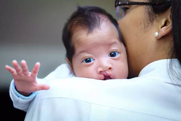
CLEFT LIP AND PALATE REPAIR
Cleft lip and Palate form during the first few weeks in pregnancy the areas of the face develop separately, including the left and right sides of the upper jaw, mouth and lips. In some cases the parts of the lip and roof of the mouth do not join properly resulting in a cleft. If the separation occurs in the upper lip, the child is said to have a cleft lip and if the separation occurs on the palate, the child is said to have cleft palate (opening on the roof of your mouth).
CLEFT LIP AND PALATE REPAIR
If both the lip and palate do not meet in the midline as supposed the child will be born with a cleft lip and palate. This condition will create a split in the lip, palate or both that will affect the normal growth and development of the child A normal developed lip and palate (roof of the mouth) is important not only for a good facial appearance but also for sucking, feeding and speech. The palate is also very important when eating, It prevents food and liquids from going up into the nose. Children born with cleft lip and palate usually need interdisciplinary management and the skills of several health care providers to correct the problems associated with the defect such as feeding, speech, hearing and psychological development. The surgeries are generally done in the hospital setting and the first surgery to repair the lip is done in the first few months of the children. If the children also presented a cleft involving the roof of the mouth the surgeries to repair the palate will be done during the children’s first few years.
SURGICAL
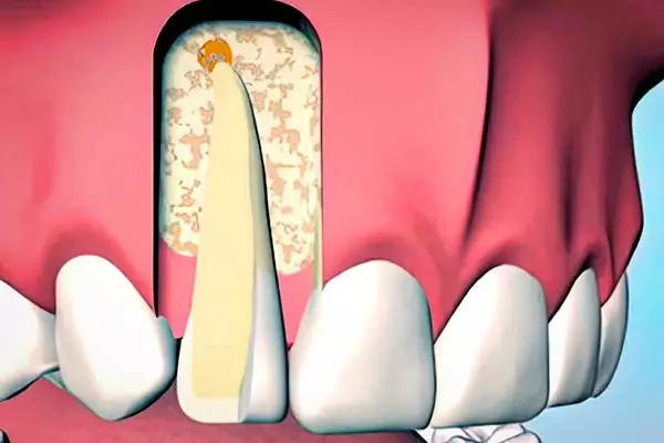
APICOECTOMY
Root canal treatments may not be sufficient to heal a tooth and surgery may be required. The most common procedure to save damaged teeth is called an apicoectomy or root-end resection. The procedure can be used to find fractures or hidden canals that do not appear on x-rays, but still manifest pain in the tooth. Affected root surfaces or the bone may also be treated during this procedure. The surgery requires an incision that is made in the gum tissue to expose the bone and the root of the tooth. The damaged tissue is then removed along with the end of the root tip a root end filling is placed to prevent reinfection of the root and the gum is sutured over. The bone naturally heals around the root and function is restored.
SURGICAL
APICOECTOMY
Root canal treatments may not be sufficient to heal a tooth and surgery may be required. The most common procedure to save damaged teeth is called an apicoectomy or root-end resection. The procedure can be used to find fractures or hidden canals that do not appear on x-rays, but still manifest pain in the tooth. Affected root surfaces or the bone may also be treated during this procedure. The surgery requires an incision that is made in the gum tissue to expose the bone and the root of the tooth. The damaged tissue is then removed along with the end of the root tip a root end filling is placed to prevent reinfection of the root and the gum is sutured over. The bone naturally heals around the root and function is restored.
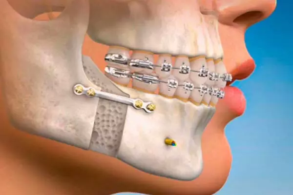
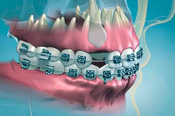
IMPACTED CANINES
The maxillary cuspid or canine (upper eyetooth) may become impacted as a result of lack of space for its proper eruption into the dental arch. The cuspid tooth represents an important role in your “bite”, they are very strong teeth for chewing and have the longest roots of any human teeth. When cuspids teeth are impacted they may be positioned on the palate (roof of the mouth) side of the dental arch next to the roots of the other anterior teeth, or they may also be located in the front of these teeth. In cases where the cuspid will not erupt spontaneously, the orthodontist and oral surgeon work together to assist the eruption of the cuspids, first the orthodontist using braces will create space for the tooth to erupt into the arch, then the patient is referred to the oral surgeon office where the gums are lifted up, the cuspid is exposed and a bracket with a gold chain is placed on the tooth so the orthodontist can complete the treatment.
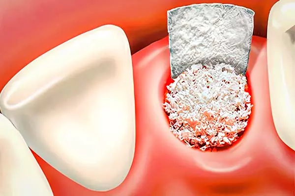
BONE GRAFTING/BONE REGENERATION
AND RECONSTRUCTION
The jawbone associated with missing teeth atrophies or is reabsorbed after the teeth are removed, this process takes a few weeks or even months after the teeth are removed. The bone of the jaws may also be lost as a result of trauma, accidents, tumors, cysts or gum disease. There may be other particular bone diseases or medications that affect normal bone function and healing. When the rate of bone loss from the jaws is quick due to any of the conditions mentioned above, the tissues around the mouth will have no support and the replacement of the teeth either with implants or other forms will be difficult.
More Details
The oral surgeons are trained to increase the amount of bone of the jaws for a foundation to replace the dentition and restore the support given by the teeth to your lips and cheeks which will give you a better aesthetic appearance. The bone used is either obtained from a tissue bank or from your own jaw bone, after a healing period of a few months the bone is ready to be restored in order to place the implants or any other form of teeth restoration.
ORAL PATHOLOGY (BIOPSY)
The lining inside the mouth is called mucosa which should be pink in color and soft. Any changes from this normal texture should be evaluated to determine is there are abnormal sores, swelling, or any white, red or pigmented (dark ,bluish, black) areas These changes can be seen any where within you mouth including lips, tongue and roof of the mouth and it may be a sign of oral cancer or a precancerous lesion. Sometimes it may be associated with swollen glands (lymphadenopathy) in the neck. Dental recall examinations are important , your dentist will examine for any of these conditions and if biopsy (removal of lesion for microscopic evaluation) is necessary you may be referred to the oral surgeon for treatment.
CLEFT LIP AND PALATE REPAIR
If both the lip and palate do not meet in the midline as supposed the child will be born with a cleft lip and palate. This condition will create a split in the lip, palate or both that will affect the normal growth and development of the child A normal developed lip and palate (roof of the mouth) is important not only for a good facial appearance but also for sucking, feeding and speech. The palate is also very important when eating, It prevents food and liquids from going up into the nose. Children born with cleft lip and palate usually need interdisciplinary management and the skills of several health care providers to correct the problems associated with the defect such as feeding, speech, hearing and psychological development. The surgeries are generally done in the hospital setting and the first surgery to repair the lip is done in the first few months of the children. If the children also presented a cleft involving the roof of the mouth the surgeries to repair the palate will be done during the children’s first few years.
BONE GRAFTING/BONE REGENERATION AND RECONSTRUCTION
The oral surgeons are trained to increase the amount of bone of the jaws for a foundation to replace the dentition and restore the support given by the teeth to your lips and cheeks which will give you a better aesthetic appearance. The bone used is either obtained from a tissue bank or from your own jaw bone, after a healing period of a few months the bone is ready to be restored in order to place the implants or any other form of teeth restoration.